
How to Choose a Laboratory Microscope in Kenya
Monocular, binocular and clinical microscopes compared for schools and diagnostic labs.
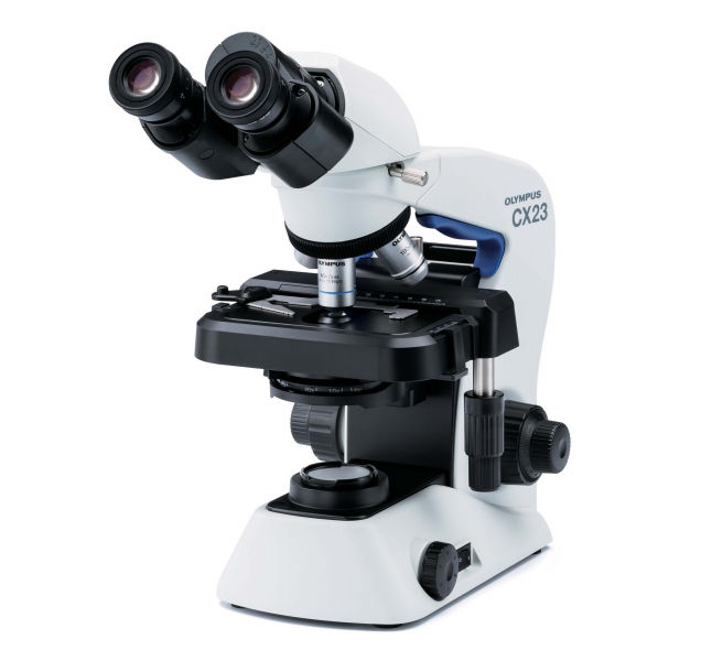
How to Choose a Laboratory Microscope in Kenya: Monocular, Binocular and Clinical Microscopes Compared
Choosing the right laboratory microscope is an important decision for schools, medical training institutions, hospitals, clinics and diagnostic laboratories in Kenya. The right microscope improves specimen visibility, supports accurate observation, makes laboratory work more comfortable and helps institutions get better value from their equipment budgets.
However, not every laboratory needs the same type of microscope. A secondary school teaching laboratory has different requirements from a university microbiology department or a clinical laboratory performing routine diagnostic investigations. Factors such as magnification, optical quality, illumination, mechanical stability, intended applications, maintenance requirements and the availability of replacement parts should all influence the purchasing decision.
Three commonly considered options are monocular microscopes, binocular microscopes and clinical laboratory microscopes. Although these categories can overlap, understanding their differences helps buyers choose equipment that matches their workload and budget.
In this guide, Zanmed Medical Supplies Ltd explains how to choose a laboratory microscope in Kenya, the specifications to compare, typical applications in educational and healthcare settings, maintenance requirements and the questions to ask before purchasing.
1. What Is a Laboratory Microscope?
A laboratory microscope is an optical instrument that magnifies small specimens, allowing users to observe structures that cannot be seen clearly with the naked eye.
In educational settings, microscopes help students study cells, tissues, microorganisms and other biological structures. In healthcare laboratories, they can support the examination of prepared slides, blood films, urine sediments, stool preparations and other specimens, depending on the diagnostic procedure and microscope configuration.
A conventional compound light microscope uses a combination of objective lenses and an eyepiece to magnify a specimen. The specimen is placed on a stage, illuminated by a light source, and brought into focus using adjustment controls.
The final image depends on several factors, including the quality of the optics, the numerical aperture of the objective and condenser, the illumination system, specimen preparation and the user's microscopy technique.
Important: High magnification does not automatically mean better image quality. A well-designed microscope with appropriate optics and illumination can provide more useful detail than a microscope offering a higher advertised magnification but poor resolution.
2. Understanding the Main Types of Laboratory Microscopes
Before comparing individual features, it is useful to understand the differences between monocular, binocular and clinical microscopes.
Monocular microscopes
A monocular microscope has one eyepiece and is viewed with one eye at a time.
It is often considered for basic biology laboratories, schools, colleges and introductory practical lessons where students are learning microscopy fundamentals.
Common features include:
Single eyepiece for observation.
Objective lenses providing different magnification levels.
A mechanical or basic specimen stage, depending on the model.
Mirror or built-in illumination, depending on the design.
Coarse and, on many models, fine focusing controls.
A relatively straightforward operating design.
Advantages: Monocular microscopes can be suitable for institutions that need multiple teaching units while working within a limited equipment budget. Their straightforward design can also make them useful for introductory practical sessions.
Limitations: Viewing through one eyepiece may be less comfortable during extended sessions. Basic models may also lack the mechanical stage, optical performance or illumination consistency required for demanding diagnostic work.
Best suited for: Basic school laboratories, introductory biology classes and educational demonstrations where the selected model meets the curriculum requirements.
Binocular microscopes
A binocular microscope has two eyepieces, allowing users to observe a specimen with both eyes.
This configuration is generally more comfortable for repeated or prolonged observation. It is commonly used in university laboratories, medical training institutions, research facilities and clinical laboratories.
Typical features include:
Two eyepieces for comfortable viewing.
Adjustable interpupillary distance on many models.
Multiple objective lenses.
Coarse and fine focusing controls.
Built-in illumination on most modern laboratory models.
Mechanical stages on many models.
Optional or model-dependent ergonomic features.
Advantages: Binocular viewing can reduce eye strain associated with keeping one eye closed and is convenient for routine laboratory work. Models with good optics, stable stages and reliable illumination are appropriate for a wide range of educational and laboratory applications.
Limitations: Binocular microscopes typically cost more than comparable basic monocular models. The quality of the optics, focusing mechanism and mechanical stage still varies considerably between products.
Best suited for: Medical training colleges, university laboratories, clinical laboratories, research facilities and institutions requiring regular or extended microscope use.
Clinical laboratory microscopes
A clinical microscope is a microscope selected and configured for healthcare laboratory work. Many conventional clinical microscopes are binocular compound light microscopes, although monocular and other configurations also exist.
The term clinical microscope describes its intended application rather than one universal optical design.
Depending on the intended tests, a clinical microscope may require:
High-quality achromatic or plan-achromatic objectives.
A mechanical stage for accurate specimen positioning.
Coarse and fine focusing controls.
An adjustable condenser.
Controlled illumination with adjustable intensity.
Suitable objectives for oil-immersion microscopy when required.
Durable construction for repeated daily use.
Compatible accessories and access to servicing.
Clinical microscopes are used for tasks such as examining stained blood films, observing urine sediments, studying parasites and performing other microscopy procedures within the laboratory's approved testing scope.
Advantages: A properly selected clinical microscope can provide the optical performance, stable specimen positioning and operational reliability needed for routine laboratory examinations.
Limitations: The appropriate configuration depends on the tests being performed. A basic clinical microscope may not be sufficient for fluorescence microscopy, phase-contrast microscopy or other specialized techniques.
Best suited for: Hospital laboratories, diagnostic centres, private laboratories, pathology departments and healthcare institutions performing microscopy-based investigations.
3. Monocular vs Binocular vs Clinical Microscopes: Comparison
The following table summarizes the main differences. Actual features and prices depend on the specific model rather than the category alone.
Feature
Monocular Microscope
Binocular Microscope
Clinical Microscope
Viewing arrangement
One eyepiece
Two eyepieces
Usually binocular; configuration varies
Typical purpose
Basic education
Education, training and routine laboratory work
Clinical and diagnostic microscopy
Viewing comfort
Suitable for shorter sessions
Generally more comfortable for prolonged use
Depends on viewing configuration and ergonomics
Optical requirements
Appropriate for intended lessons
Depends on model and objectives
Must meet the requirements of the diagnostic procedure
Illumination
Mirror or built-in light, depending on model
Usually built-in illumination
Controlled illumination suited to clinical specimens
Mechanical stage
Model-dependent
Common on laboratory models
Generally desirable for routine clinical work
Focusing
Coarse; fine focus may also be included
Usually coarse and fine
Precise coarse and fine focusing is important
Oil-immersion capability
Depends on included objectives
Depends on included objectives
Often required for specific applications, such as certain blood-film examinations
Typical purchasing priority
Affordability and teaching suitability
Comfort, optics and durability
Diagnostic suitability, optical performance and reliability
Common users
Schools and introductory laboratories
Colleges, universities and laboratories
Hospitals and diagnostic laboratories
A binocular microscope can also be a clinical microscope. These terms are not mutually exclusive: one describes the viewing arrangement, while the other describes the intended use.
4. What Magnification Do You Need?
Magnification is one of the first specifications buyers compare. However, selecting a microscope based only on its maximum advertised magnification is a common purchasing mistake.
A compound microscope's total magnification is calculated by multiplying the magnification of the eyepiece by the magnification of the objective lens.
For example:
Eyepiece
Objective
Total Magnification
10×
4×
40×
10×
10×
100×
10×
40×
400×
10×
100× oil immersion
1,000×
A microscope with a 10× eyepiece and a 40× objective therefore provides 400× total magnification.
40× magnification
This is useful for locating specimens and observing larger structures. It is commonly used as a starting point when scanning a prepared slide.
100× magnification
This provides greater detail and is useful for examining many prepared biological specimens, depending on the objective and the specimen.
400× magnification
This is a common working magnification for educational biology, cell observation and many routine laboratory microscopy procedures.
1,000× magnification
This is typically achieved using a 100× oil-immersion objective and a 10× eyepiece. It is useful for specific applications, including detailed examination of stained blood films, when the microscope's optics and the procedure support it.
Oil immersion requires the appropriate immersion oil, objective and technique. It should not be used with an objective that is not designed for immersion.
Purchasing recommendation: For many general biology and routine clinical microscopy applications, a microscope with 4×, 10× and 40× objectives is a practical starting point. If the laboratory performs procedures requiring oil immersion, confirm whether a 100× oil-immersion objective is included or available.
5. Optical Quality: Resolution Matters More Than Maximum Magnification
Two microscopes can offer the same nominal magnification but produce noticeably different images.
This difference often comes down to optical quality, numerical aperture, lens design, illumination and alignment.
Achromatic objectives
Achromatic objectives correct certain optical colour errors and are commonly used in educational and routine laboratory microscopes.
They are suitable for many general microscopy applications when properly matched with the rest of the optical system.
Plan-achromatic objectives
Plan-achromatic objectives are designed to provide a flatter field of view, helping keep more of the specimen in focus across the image.
They can be particularly useful when examining specimens across a wider field, including routine laboratory slide examination.
Numerical aperture
Numerical aperture, commonly abbreviated as NA, is an important indicator of an objective's light-gathering ability and potential resolving performance.
A higher NA can help resolve finer detail when the optical system and illumination are properly matched.
Eyepiece field number
The field number helps determine how much of the specimen is visible through the eyepiece. A wider field can make scanning and positioning specimens more convenient.
What should buyers choose? Schools teaching basic microscopy may be well served by good-quality achromatic objectives. Clinical laboratories performing frequent slide examinations may benefit from plan-achromatic objectives, depending on the workload and budget.
Do not pay a premium for a particular optical label without checking the actual objective specifications, image quality and suitability for your intended applications.
6. Illumination: LED or Halogen?
The illumination system affects specimen visibility, contrast, convenience and operating costs.
LED illumination
LED illumination is common in modern laboratory microscopes.
Potential benefits include:
Lower power consumption.
Long service life.
Relatively low heat generation.
Adjustable brightness on many models.
Convenient operation in classrooms and clinical laboratories.
Some LED microscopes can operate using rechargeable batteries or external power arrangements, depending on the model.
Halogen illumination
Halogen lighting has traditionally been used in many laboratory microscopes.
Potential benefits include:
A familiar light quality for experienced users.
Adjustable brightness on many models.
Compatibility with established microscope designs.
Halogen lamps generally produce more heat and require periodic replacement. The availability and cost of replacement lamps should be considered when purchasing older or established microscope models.
Which is better?
For many new school and routine laboratory installations, an LED microscope is an attractive choice because of its efficiency and low-maintenance lighting.
However, a well-maintained halogen microscope can still perform effectively. The better choice depends on optical performance, user preference, power requirements, replacement-part availability and total cost of ownership.
7. Mechanical Stage and Focusing Controls
The stage holds the specimen slide while the focusing mechanism brings the specimen into clear view.
These features have a direct impact on the microscope's usability.
Mechanical stage
A mechanical stage allows the user to move the slide in controlled horizontal and vertical directions.
It makes it easier to scan specimens systematically, return to an area of interest and position a slide accurately.
For routine clinical microscopy, a mechanical stage with smooth controls is generally preferable to a basic stage that requires the slide to be moved entirely by hand.
Coarse focusing
The coarse-focus control moves the stage or optical assembly relatively quickly to bring the specimen into approximate focus.
Fine focusing
The fine-focus control makes smaller adjustments to sharpen the image. It is particularly important at higher magnifications, where small movements can significantly affect focus.
Rackless stage design
Some microscopes use rackless stage designs that reduce exposed protruding mechanical components. This may improve safety and durability in teaching environments.
When comparing models, test the stage movement and focusing controls if possible. They should operate smoothly without excessive looseness or abrupt movement.
8. Choosing a Microscope for Schools in Kenya
Schools need microscopes that support practical learning while remaining affordable to acquire and maintain.
The right choice depends on the number of students, curriculum, class size, frequency of practical lessons and available laboratory budget.
Recommended considerations
1. Ease of use
Choose a microscope with straightforward controls and a stable base so students can learn focusing, slide positioning and illumination without unnecessary difficulty.
2. Suitable magnification
For introductory biology, 40×, 100× and 400× total magnification will cover many common learning activities.
3. Durable construction
The microscope should withstand routine classroom use. A stable frame, robust stage and reliable focusing mechanism are valuable.
4. Illumination
Built-in LED illumination is convenient for many modern school laboratories, particularly where consistent lighting and lower maintenance are priorities.
5. Number of units
Calculate the number of microscopes required for the expected student groups. A practical arrangement may involve several students sharing one microscope during a lesson, but the class format and curriculum should determine the final number.
6. Replacement parts
Ask whether the supplier can support the microscope with replacement eyepieces, objectives, lamps or LEDs, fuses and other serviceable components.
Monocular or binocular for schools?
A monocular microscope may be suitable for introductory lessons and budget-conscious purchases.
A binocular microscope may be worth the additional cost where students undertake frequent practical sessions or where the institution wants a more comfortable viewing experience.
For advanced biology and medical training, the required optical performance and teaching objectives should guide the selection rather than price alone.
9. Choosing a Microscope for Hospitals and Diagnostic Laboratories
Clinical laboratories need microscopes selected around their actual diagnostic procedures, workload and quality requirements.
A microscope that is adequate for a school practical lesson may not provide the ergonomics, optics or mechanical stability required for a busy diagnostic laboratory.
Routine blood-film examination
Blood-film microscopy may require a good-quality 100× oil-immersion objective, appropriate illumination and stable fine focusing.
The laboratory should confirm that the microscope supports its established blood-film examination procedure.
Urine microscopy
Urine sediment examination requires appropriate objectives, reliable focusing and sufficient illumination. The exact magnification and technique depend on the laboratory's procedure.
Parasitology
Microscopy may be used for examining specimens for parasites, including malaria parasites on appropriately prepared blood films. The required microscope configuration depends on the examination and staining method.
Stool microscopy
Microscopy may support the examination of prepared stool specimens for parasites and other structures, according to the laboratory's testing procedures.
Microbiology and educational laboratory work
General compound microscopes can support many microscopy teaching and observation tasks. Specialized applications may require different optical methods or additional equipment.
Key requirements for clinical buyers
Suitable objective lenses for the tests performed.
Clear optics with good contrast and resolution.
Stable, smooth mechanical stage movement.
Coarse and fine focusing controls.
Adjustable condenser and illumination.
Comfortable eyepiece arrangement for prolonged use.
Durable construction for routine daily work.
Access to technical support and replacement parts.
Clinical use also requires trained personnel, suitable specimen preparation, validated procedures and adherence to the facility's quality-management requirements. The microscope alone does not guarantee an accurate diagnosis.
10. Understanding the Olympus CX21 Microscope
The Olympus CX21 is an established biological microscope design that may be considered by educational institutions, medical training colleges and laboratories looking for a conventional compound microscope.
Depending on the specific configuration and condition, models in this family can offer features such as binocular viewing, mechanical specimen positioning, coarse and fine focusing, and halogen illumination.
When evaluating an Olympus CX21, or any comparable microscope, confirm the exact configuration being supplied.
Check the following:
Whether the microscope is new, refurbished or used.
The condition of the eyepieces and objective lenses.
Whether the supplied objectives are 4×, 10× and 40×.
Whether a 100× oil-immersion objective is included.
The condition of the condenser and iris diaphragm.
The performance of the illumination system.
Whether the mechanical stage moves smoothly.
Whether the fine-focus control operates correctly.
Whether the correct power supply and replacement lamps are available.
Whether servicing and spare parts can be obtained locally.
Some established microscope designs use halogen illumination rather than LED illumination. This is not automatically a disadvantage, but lamp availability and ongoing maintenance should form part of the purchasing decision.
Buyer tip: Never assume that every Olympus CX21 listing contains identical accessories or specifications. Verify the exact unit, included objectives, condition and warranty before committing to a purchase.
11. How Much Does a Laboratory Microscope Cost in Kenya?
Laboratory microscope prices in Kenya vary by type, optical specification, brand, condition, accessories and supplier.
A basic monocular teaching microscope will generally occupy a different price category from a binocular clinical microscope with higher-grade objectives and a mechanical stage.
Instead of choosing solely by the lowest quoted price, compare the full specification and the total cost of ownership.
Factors that affect microscope prices
Optical system: Objective quality and optical design can significantly affect image quality and price.
Viewing configuration: Monocular and binocular models may differ in price, although the exact difference depends on their other specifications.
Illumination: LED and halogen systems have different purchase and maintenance considerations.
Mechanical stage: A precision mechanical stage may add to the cost but can improve usability.
Brand and model: Established brands and specialized configurations may command higher prices.
Condition: New, refurbished and used microscopes should not be compared as though they carry the same risk or warranty.
Accessories: Extra objectives, spare eyepieces, immersion oil, dust covers and other accessories can affect the final quotation.
After-sales support: Warranty coverage, servicing and access to replacement parts contribute to long-term value.
How to compare quotations
Ask suppliers to quote on a like-for-like basis. A useful comparison should identify:
Manufacturer and exact model.
Monocular or binocular configuration.
Objective magnifications and types.
Eyepiece magnification and field number.
Illumination type.
Mechanical or basic stage.
Condenser specification.
Included accessories.
Warranty period and coverage.
Delivery and installation charges, where applicable.
Availability of servicing and spare parts.
Applicable taxes and final delivered price.
For budgeting purposes, obtain current quotations rather than relying on old online prices. This is particularly important when purchasing multiple microscopes for schools, universities, hospitals or institutional tenders.
12. How to Calculate the Total Cost of Ownership
The initial purchase price is only one part of the cost of owning a microscope.
A more useful comparison considers the expected cost of using and maintaining the equipment over several years.
A simple budgeting formula is:
Total cost of ownership = Purchase price + Maintenance + Consumables + Repairs + Replacement parts
For example, consider a hypothetical microscope purchased for KSh 30,000.
Cost Item
Illustrative Amount
Initial purchase
KSh 30,000
Planned maintenance over 3 years
KSh 3,000
Replacement lamp or minor parts
KSh 2,000
Cleaning and related supplies
KSh 1,500
Estimated 3-year total
KSh 36,500
These figures are illustrative only, not a current supplier quotation or a forecast of actual repair costs.
If the microscope is used for three years, the estimated cost averages approximately KSh 12,167 per year, before considering financing, downtime or any resale value.
When comparing alternatives, calculate the same costs over the same period. A microscope with a higher purchase price may offer better value if it provides greater durability, lower maintenance requirements or more dependable local service.
13. Microscope Maintenance: How to Protect Your Investment
Regular maintenance helps preserve optical performance, reduce avoidable damage and extend the useful life of a microscope.
After each use
Remove the specimen slide carefully.
Clean immersion oil from the 100× objective using the recommended lens-cleaning materials.
Switch off the illumination and disconnect the power when appropriate.
Return the microscope to a safe, stable position.
Cover it with a suitable dust cover after it has cooled, where applicable.
Routine cleaning
Use lens paper or manufacturer-approved optical cleaning materials on lenses. Ordinary tissue, rough cloth and unsuitable solvents can scratch or damage optical coatings.
Clean external surfaces using a method appropriate to the manufacturer's instructions and the laboratory's infection-control procedures.
Storage
Store microscopes in a clean, dry and secure environment. Protect them from dust, excessive humidity, direct sunlight, accidental impact and unauthorized handling.
In humid environments, appropriate storage practices can help reduce the risk of fungal growth on optical components.
Periodic inspection
Check the following regularly:
Eyepieces and objective lenses.
Stage movement and slide holder.
Coarse and fine focusing controls.
Condenser and iris diaphragm.
Illumination and electrical connections.
Mechanical stability.
Power cable and switch.
Arrange professional servicing when optical alignment, focusing or electrical operation becomes unreliable.
14. Common Mistakes to Avoid When Buying a Microscope
Buying based on magnification alone
A high magnification figure does not guarantee clear detail. Check optical quality and resolution.
Choosing the wrong microscope for the application
A basic educational microscope may not be suitable for a laboratory performing routine diagnostic examinations.
Ignoring the objective lenses
Confirm which objectives are included. A microscope described as capable of 1,000× magnification may require an additional 100× objective and immersion oil.
Overlooking illumination requirements
Consider brightness control, light consistency, lamp availability and power requirements.
Ignoring ergonomics
For frequent use, binocular viewing, comfortable controls and a stable stage can improve the working experience.
Failing to check after-sales support
Before purchasing, establish whether technical assistance, replacement parts and warranty service are available.
Assuming all clinical microscopes are equivalent
Different tests may require different optical specifications. Select equipment based on the laboratory's actual procedures.
Comparing prices without comparing specifications
A lower quotation may exclude important accessories or offer a shorter warranty. Compare the complete package rather than the headline price.
15. Microscope Purchasing Checklist for Kenyan Institutions
Use this checklist when preparing a laboratory equipment order or requesting a supplier quotation.
Define the intended applications.
Determine the number of microscopes required.
Select monocular or binocular viewing as appropriate.
Confirm the required objective magnifications.
Verify whether oil-immersion capability is needed.
Check objective quality and optical specifications.
Confirm illumination type and brightness adjustment.
Check whether a mechanical stage is required.
Verify condenser and focusing specifications.
Confirm the microscope's physical condition.
Request warranty and servicing information.
Confirm the availability of replacement parts.
Check included accessories.
Compare final delivered prices and applicable taxes.
Confirm delivery arrangements and any installation or training requirements.
For public institutions and larger organizations, ensure the proposed equipment also meets the institution's technical specifications, procurement procedures and any applicable regulatory or quality requirements.
16. Where to Buy Laboratory Microscopes in Kenya
When sourcing a microscope, choose a supplier that can clearly explain the available models, technical specifications, included accessories and after-sales support.
For schools and training institutions, prioritize a microscope that supports the curriculum and can withstand repeated student use. For diagnostic laboratories, prioritize optical performance, the required objectives, reliable focusing and serviceability.
Zanmed Medical Supplies Ltd supplies medical and laboratory equipment for healthcare providers, educational institutions and other organizations in Kenya.
Whether you need a microscope for a school biology laboratory, medical training facility, hospital laboratory or diagnostic centre, request a quotation based on your intended applications, preferred configuration and budget.
Before placing an order, confirm product availability, the exact model, included objectives, warranty, delivery arrangements and current pricing.
Frequently Asked Questions
Which microscope is best for a school laboratory in Kenya?
A good-quality monocular or binocular compound microscope can suit a school laboratory. The choice depends on the curriculum, frequency of practical sessions, number of students and available budget. Models offering 40×, 100× and 400× total magnification cover many introductory biology activities.
Which microscope is best for a clinical laboratory?
A binocular compound microscope with good optics, a mechanical stage, coarse and fine focusing, suitable illumination and the required objectives is a common choice for routine clinical microscopy. The final specification should match the laboratory's actual tests.
What is the difference between a monocular and binocular microscope?
A monocular microscope has one eyepiece and is viewed with one eye. A binocular microscope has two eyepieces and allows viewing with both eyes, which many users find more comfortable during prolonged observation.
Is a binocular microscope suitable for medical laboratory work?
Yes. Binocular microscopes are widely suitable for medical laboratory work when their optics, objectives, illumination, stage and other features meet the requirements of the intended examination.
Is 1,000× magnification necessary?
Not for every application. Many educational tasks and routine examinations use lower magnifications. A 100× oil-immersion objective, typically providing 1,000× total magnification with a 10× eyepiece, is required for certain procedures, including some stained blood-film examinations.
Is LED illumination better than halogen?
LED illumination generally offers lower power consumption, long service life and less heat. Halogen illumination can also provide satisfactory performance. Consider the microscope's image quality, replacement-part availability and intended use.
What should I check before buying an Olympus CX21 microscope?
Confirm its condition, exact configuration, objective lenses, illumination system, condenser, mechanical stage, focusing operation, warranty and availability of servicing and spare parts. Do not assume that every unit is supplied with the same accessories.
How often should a laboratory microscope be serviced?
The frequency depends on the manufacturer's recommendations, usage and laboratory environment. Routine cleaning and inspection should be performed regularly, with professional servicing arranged as recommended or whenever optical, mechanical or electrical problems arise.
Can a laboratory microscope be used to diagnose malaria?
Microscopy can support malaria diagnosis when appropriate blood films are prepared and examined by trained personnel using suitable equipment and validated procedures. A microscope alone does not establish a diagnosis.
Conclusion
Choosing the right laboratory microscope in Kenya requires more than comparing magnification figures or selecting the cheapest available model. Buyers should consider the intended application, optical quality, objective lenses, illumination, stage design, focusing controls, durability, maintenance and availability of technical support.
For schools, a reliable monocular or binocular microscope may provide the functionality needed for biology lessons. For hospitals and diagnostic laboratories, a well-specified clinical microscope can support routine microscopy when matched to the required procedures.
By comparing complete specifications and total ownership costs, Kenyan institutions can invest in laboratory microscopes that meet their needs, support dependable laboratory work and provide long-term value.
Looking for laboratory microscopes in Kenya? Contact Zanmed Medical Supplies Ltd to discuss your laboratory requirements and request a quotation for suitable microscope options.